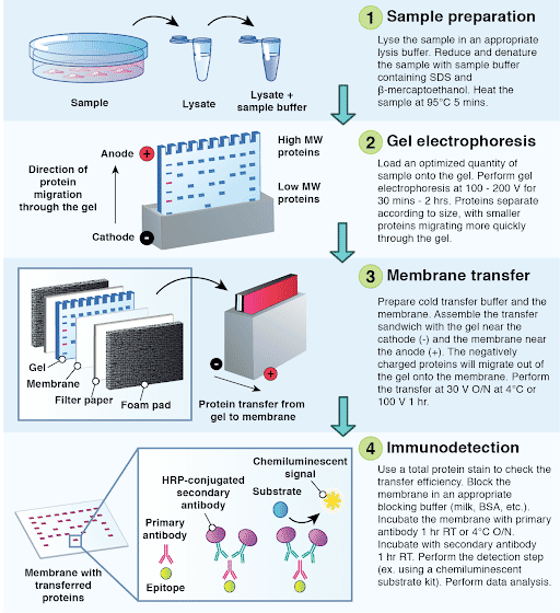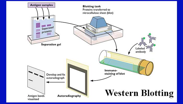

The albumin concentration served as comparison parameter among the single animals and groups. After washing with PBST (PBS, 0.05% Tween-20), the membranes were incubated for 1 h with HRP-conjugated donkey anti-chicken IgG (Dianova, Hamburg, Germany) or donkey anti-mouse IgG (Jackson ImmunoResearch, Cambridge, UK) at a dilution of 1:10,000 and were finally developed using Western Lightning Plus-ECL kit (PerkinElmer, Waltham, MA, USA) with the ChemiDoc Touch imaging system (Bio-Rad, Hercules, CA, USA). After blocking for 30 min with a blocking buffer (5% nonfat dry milk, PBS, 0.05% Tween-20), the membranes were incubated with the primary antibody at 4 ☌ overnight at the following dilutions: anti-albumin mAb 1:1000 (ab106582, Abcam, Cambridge, UK) and anti-actin mAb 1:1,000,000 (#A5441, Merck, Darmstadt, Germany).

20 µg of the total protein was loaded on the gel, electrophoresed and transferred to a nitrocellulose membrane. The lysates were mixed with a 2 × SDS-PAGE loading buffer (final concentration: 60 mM Tris, pH 6.8, 10% beta-mercaptoethanol, 5% SDS, 10% glycerol) at 95 ☌ for 10 min.

After centrifugation at 15,000× g for 30 min at 4 ☌, supernatants were used for a bicinchoninic acid (BCA) protein assay and subsequent WB analysis. Dissected cortices and basal ganglia from mouse brains were homogenized with a RIPA buffer (25 mM Tris pH 7.4, 150 mM NaCl, 1% NP-40, 0.1% SDS) containing 4% proteinase inhibitor (cOmplete protease inhibitor cocktail, Sigma Aldrich, Germany) and sonified for 10 s. This method was used in the original publication out of which the results were taken for correlation. With researchers expected to adhere to the 3R-principle to replace, reduce and refine animal experiments, alternatives are necessary. Furthermore, growing evidence revealed the toxicity of Evans Blue, with an increased periprocedural mortality of the animals. Yet after the aforementioned measurements, the dyed tissue is no longer suitable for further histological or biochemical investigations. It is posthumously assessed either qualitatively, by a simple visualization of blue discoloration within the sectioned brain tissue, or quantitatively, by incubating the extracted tissue in formamide and measuring the dye concentration with ultraviolet spectrophotometry. When it comes to the determination of the BBB function post mortem, a mainstay has long been the Evans Blue dye technique, in which the blue azo dye, binding to serum albumin, is injected in the jugular or tail vein of the living murine animals. Commonly used isoflurane narcosis, in particular, is associated with neuroprotective bystander effects. This technology, though, requires further anesthesia, which might interfere with the superior aims of the respective studies. Another well-established live imaging technique to examine BBB integrity is in-vivo two-photon microscopy. To the first group belong the undoubtedly informative, but at the same time labor-intensive, equipment- and infrastructure-dependent as well as expensive live imaging technologies like magnetic resonance imaging (MRI) or computed tomography (CT) with perfusion-sensitive weightings or as single photon emission CT (SPECT). Various methods to measure the degree of BBB disruption in murine models exist, both in vivo and ex vivo.

This is the reason why researchers, acting within the scope of BBB-compromising diseases in general or within the environment of experimental strokes in special, require convenient and reliable techniques to characterize the BBB integrity.


 0 kommentar(er)
